what does the left subclavian artery branch to
| Subclavian artery | |
|---|---|
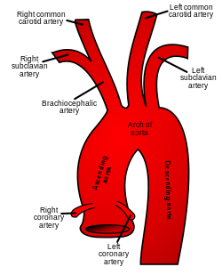 Schematic of the proximal aorta and its branches. The left subclavian artery is the fifth co-operative of the aorta and the tertiary co-operative from the arch of the aorta. The correct subclavian artery arises from the brachiocephalic artery and its branches. (Right subclavian is at upper left, and left subclavian is at upper right.) | |
| Details | |
| Source | aortic arch (left) brachiocephalic (right) |
| Branches | vertebral artery internal thoracic avenue thyrocervical torso costocervical trunk dorsal scapular artery (mostly) |
| Vein | subclavian vein |
| Identifiers | |
| Latin | arteria subclavia |
| MeSH | D013348 |
| TA98 | A12.2.08.001 |
| TA2 | 4537 |
| FMA | 3951 |
| Anatomical terminology [edit on Wikidata] | |
In man anatomy, the subclavian arteries are paired major arteries of the upper thorax, below the clavicle. They receive blood from the aortic curvation. The left subclavian avenue supplies blood to the left arm and the right subclavian artery supplies blood to the correct arm, with some branches supplying the caput and thorax. On the left side of the torso, the subclavian comes direct off the aortic arch, while on the right side information technology arises from the relatively brusk brachiocephalic artery when information technology bifurcates into the subclavian and the right common carotid avenue.
The usual branches of the subclavian on both sides of the body are the vertebral artery, the internal thoracic artery, the thyrocervical trunk, the costocervical body and the dorsal scapular artery, which may co-operative off the transverse cervical artery, which is a branch of the thyrocervical trunk. The subclavian becomes the axillary artery at the lateral border of the get-go rib.
Structure [edit]
From its origin, the subclavian artery travels laterally, passing between anterior and middle scalene muscles, with the anterior scalene (scalenus anterior) on its anterior side and the center scalene (scalenus medius) on its posterior. This is in contrast to the subclavian vein, which travels inductive to the scalenus anterior. As the subclavian avenue crosses the lateral border of the first rib, it becomes the axillary artery.[1] [2]
On the correct side the subclavian artery arises from the brachiocephalic (innominate) artery behind the right sternoclavicular articulation; on the left side information technology springs from the arch of the aorta.[1] [3] The two vessels, therefore, in the first function of their form, differ in length, direction, and relation with neighboring structures.[iv] The left subclavian artery is around 9 cm long in adults, while the right subclavian avenue is around 6 cm long.[four] Both take a width of ix-12 mm.[4]
Parts [edit]
In club to facilitate the clarification, each subclavian artery is divided into 3 parts:
- The first part, also known as the prescalene part,[4] extends from the origin of the vessel to the medial border of the scalenus anterior muscle.[1]
- The second part, also known as the scalene part,[4] lies behind the scalenus anterior muscle.[1]
- The 3rd office, also known as the postscalene function,[iv] extends from the lateral margin of the musculus to the outer border of the first rib, where it becomes the axillary artery.[1] [2]
The get-go portions of the 2 vessels require split descriptions; the second and third parts of the two arteries are practically alike.
Offset part [edit]
Correct subclavian artery [edit]
The first function of the right subclavian artery arises from the brachiocephalic trunk, backside the upper part of the right sternoclavicular articulation.[1] [3] It passes upward and lateralward to the medial margin of the scalenus anterior muscle. Information technology ascends a footling above the medial part of the clavicle.[four]
It is covered, in front, past the integument, superficial fascia, the platysma muscle, deep fascia, the clavicular origin of the sternocleidomastoid muscle, the sternohyoid musculus, and the sternothyroid muscle, and another layer of the deep fascia. It is crossed by the internal jugular vein and the vertebral vein, by the vagus nerve and the cardiac branches of the vagus and sympathetic, and past the subclavian loop of the sympathetic trunk which forms a ring around the vessel. The inductive jugular vein is directed laterally in front of the avenue, but is separated from it past the sternohyoid muscle and the sternothyreoid muscle. Below and behind the artery is the pleura, which separates it from the apex of the lung.[4] Behind the artery is the sympathetic trunk, the longus colli muscle and the first thoracic vertebra (T1). The right recurrent laryngeal nervus winds around the lower and back part of the vessel.[iv]
Left subclavian artery [edit]
The start part of the left subclavian avenue arises from the aortic arch, behind the left common carotid avenue, and at the level of the 4th thoracic vertebra.[1] [3] It ascends in the superior mediastinal cavity to the root of the neck, and then arches lateralward to the medial border of the scalenus inductive muscle.
It is in relation, in front, with the vagus nerve, the cardiac nerves, and the phrenic fretfulness, which lie parallel with it, the left common carotid artery, left internal jugular and vertebral veins, and the start of the left innominate vein. It is covered by the sternothyroid muscle, the sternohyoid muscle, and the sternocleidomastoid muscle. Backside, it is in relation with the esophagus, thoracic duct, left recurrent laryngeal nerve, inferior cervical ganglion of the sympathetic trunk, and the longus colli muscle; higher up, notwithstanding, the esophagus and thoracic duct lie to its right side; the latter ultimately arching over the vessel to bring together the bending of matrimony between the subclavian and internal jugular veins. Medial to it are the esophagus, trachea, thoracic duct, and left recurrent laryngeal nervus. Lateral to it are the left pleura and lung.[4]
2nd part [edit]
The second portion of the subclavian artery lies behind the scalenus inductive muscle and in front of the scalenus medius muscle.[3] [4] It is very brusk, and forms the highest function of the curvation described past the vessel.
In front, it is covered past the skin, the superficial fascia, the platysma muscle, the deep cervical fascia, the sternocleidomastoid muscle, and the scalenus anterior muscle. On the correct side of the neck, the phrenic nervus is separated from the 2d part of the artery by the scalenus anterior muscle, while on the left side information technology crosses the first part of the artery close to the medial border of the musculus. Behind the artery are the pleura and the scalenus medius muscle. Higher up the artery is the brachial plexus.[4] Below the avenue is the pleura.[4] The subclavian vein lies beneath and anterior to the artery, separated from it past the scalenus anterior musculus.[four]
3rd part [edit]
The 3rd portion of the subclavian artery runs down and lateralward from the lateral margin of the scalenus anterior muscle to the outer edge of the first rib, where it becomes the axillary avenue.[i] [2] This is the most superficial portion of the vessel, and is contained in the subclavian triangle.
It is covered, in front, by the skin, the superficial fascia, the platysma musculus, the supraclavicular nerves, and the deep cervical fascia.[4] The external jugular vein crosses its medial part and receives the transverse scapular, transverse cervical, and anterior jugular veins, which frequently course a plexus in front of the avenue. Behind the veins, the nervus to the Subclavius descends in front of the avenue. The terminal part of the avenue lies behind the clavicle and the Subclavius and is crossed by the transverse scapular vessels. The subclavian vein is in front of and at a slightly lower level than the artery. Behind, it lies on the lowest torso of the brachial plexus, which intervenes between it and the scalenus medius muscle. To a higher place and to its lateral side are the upper trunks of the brachial plexus and the omohyoid muscle. Beneath, it rests on the upper surface of the first rib.
Branches [edit]
The subclavian arteries give off five major arteries each: the vertebral artery, the internal thoracic artery, the thyrocervical trunk, the costocervical trunk, and the dorsal scapular artery.[1] [4]
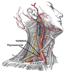
Superficial dissection of the right side of the neck, showing the carotid and subclavian arteries. Branch of vertebral artery and thyrocervical trunk is labeled. Internal thoracic avenue branches from same segment, but inferiorily, and is therefore non visible.
| Role | Branches | Course |
|---|---|---|
| Beginning role From its origin to the medial border of scalenus anterior | Vertebral artery | Runs cranially in the transverse foramina of the cervical vertebrae,[ane] joins the vertebral avenue on the contralateral side, forming the basilar artery and joins the circle of Willis. |
| Internal thoracic artery | Runs caudally behind the ribs, giving off anterior intercostal branches, perforating vessels to the chest and terminating in the superior epigastric artery and the musculophrenic artery.[1] | |
| Thyrocervical trunk | Very short. Divides into inferior thyroid artery, suprascapular artery and transverse cervical artery (also called cervicodorsal trunk).[one] | |
| Second part Lying backside scalenus inductive | Costocervical trunk | Splits into superior intercostal artery and deep cervical avenue.[1] |
| Third part Between the lateral border of scalenus inductive and the outer border of the first rib | Dorsal scapular avenue | From either second or third part.[1] Passes backwards to supply levator scapulae and rhomboids. |
Development [edit]
Embryologically, the left subclavian simply arises from the left seventh intersegmental artery,[v] while the right subclavian arises, proximal to distal:
- right quaternary aortic arch
- right dorsal aorta
- correct 7th intersegmental artery
Substantially, the 4th aortic arch and dorsal aorta form the aortic arch on the left, but since the right dorsal aorta regresses distal to the right 7th intersegmental artery, on the right they form the proximal portion of the subclavian artery. Since the left subclavian is and then a tributary of the left common carotid, they tin be thought of as arising from the brachiocephalic trunk.
Variation [edit]
The subclavian arteries vary in their origin, their course, and the height to which they rising in the cervix.
The origin of the correct subclavian from the innominate takes identify, in some cases, above the sternoclavicular articulation, and occasionally, just less often, beneath that articulation. The avenue may ascend as a carve up trunk from the curvation of the aorta, and in such cases it may be either the first, second, 3rd, or even the last branch derived from that vessel; in the majority, however, it is the offset or concluding, rarely the 2d or third. When information technology is the first co-operative, it occupies the ordinary position of the innominate artery; when the second or third, it gains its usual position by passing behind the right carotid; and when the last co-operative, it arises from the left extremity of the arch, and passes obliquely toward the right side, unremarkably behind the trachea, esophagus, and right carotid, sometimes between the esophagus and trachea, to the upper edge of the first rib, whence it follows its ordinary form. In very rare instances, this vessel arises from the thoracic aorta, as low downwards every bit the 4th thoracic vertebra. Occasionally, it perforates the Scalenus inductive; more rarely information technology passes in front of that musculus. Sometimes the subclavian vein passes with the artery behind the Scalenus inductive. The artery may arise every bit loftier every bit 4 cm. above the clavicle, or any intermediate point between this and the upper border of the bone, the correct subclavian commonly ascending higher than the left.
The left subclavian is occasionally joined at its origin with the left mutual carotid artery, forming a left brachiocephalic body.
The left subclavian artery is more deeply placed than the right in the get-go part of its grade, and, equally a rule, does non reach quite as high a level in the neck. The posterior border of the Sternocleidomastoideus corresponds pretty closely to the lateral edge of the Scalenus anterior, so that the tertiary portion of the artery, the part most attainable for functioning, lies immediately lateral to the posterior border of the Sternocleidomastoideus.
Some authors describe the subclavian artery as arising from the 7th intersegmental artery.
Part [edit]
The subclavian arteries deport most of the blood that supplies the artillery.[3] It besides supplies some blood to the neck and brain.[3]
Clinical significance [edit]
Pinch of the subclavian avenue can cause thoracic outlet syndrome (TOS).[half dozen]
The subclavian arteries tin exist vulnerable to aneurysm.[half-dozen]
Subclavian steal syndrome occurs when there is occlusion or stenosis of the subclavian avenue at a betoken earlier the branching of the vertebral avenue.[7] This tin can cause claret to flow the wrong way through the vertebral artery into the distal subclavian avenue, allowed by the reduced pressure.[7] [8]
The subclavian arteries are relatively superficial, and can exist seen using ultrasound.[8]
Aberrant right subclavian artery is a condition where the right subclavian artery arises on the arch of aorta distal to the left subclavian artery, instead of right brachocephalic torso. This condition occurs in about 0.iv to 1.8% of the general population. The abnormal right subclavian artery than has to cross behind the oesophagus to achieve the right side of the body. Bulk of those having the condition are asymptomatic, just some may be presented with difficulty in breathing, difficulty in swallowing, grating noise during breathing, and chest pain. This condition has to be ruled out during procedures relating to oesophagus because injury to this artery during the procedure tin can cause massive bleeding.[9]
Boosted images [edit]
-

Side of cervix, showing chief surface markings.
-

Magnetic Resonance Angiography
-

Right subclavian avenue
-
Brachial plexus and subclavian artery
Run into also [edit]
- Abnormal subclavian avenue
- Subclavian steal syndrome
- Thoracic outlet syndrome
References [edit]
![]() This article incorporates text in the public domain from folio 575 of the 20th edition of Gray's Anatomy (1918)
This article incorporates text in the public domain from folio 575 of the 20th edition of Gray's Anatomy (1918)
- ^ a b c d due east f m h i j k l m n Valji, Karim, ed. (2006-01-01), "Chapter 7 - Upper Extremity Arteries", Vascular and Interventional Radiology (2nd Edition), Philadelphia: West.B. Saunders, pp. 182–203, doi:10.1016/B978-0-7216-0621-vii.50012-3, ISBN978-0-7216-0621-7 , retrieved 2021-01-05
- ^ a b c Crystal, George J.; Assaad, Sherif I.; Heerdt, Paul M. (2019-01-01), Hemmings, Hugh C.; Egan, Talmage D. (eds.), "24 - Cardiovascular Physiology: Integrative Function", Pharmacology and Physiology for Anesthesia (Second Edition), Philadelphia: Elsevier, pp. 473–519, doi:10.1016/b978-0-323-48110-6.00024-vii, ISBN978-0-323-48110-half-dozen, S2CID 87519432, retrieved 2021-01-05
- ^ a b c d e f Woodward, Paula J.; Griffith, James F.; Antonio, Gregory E.; Ahuja, Anil T., eds. (2018-01-01), "Lower Cervical Level and Supraclavicular Fossa", Imaging Anatomy: Ultrasound (2nd Edition), Elsevier, pp. 124–129, doi:10.1016/b978-0-323-54800-7.50019-two, ISBN978-0-323-54800-7 , retrieved 2021-01-05
- ^ a b c d e f one thousand h i j m 50 m n o p Barral, Jean-Pierre; Croibier, Alain (2011-01-01), Barral, Jean-Pierre; Croibier, Alain (eds.), "11 - The subclavian arteries", Visceral Vascular Manipulations, Oxford: Churchill Livingstone, pp. 110–116, doi:x.1016/b978-0-7020-4351-two.00011-ix, ISBN978-0-7020-4351-2 , retrieved 2021-01-05
- ^ "Aortic arches". www.embryology.ch . Retrieved 12 July 2016.
- ^ a b Rigberg, David; Freischlag, Julie (2009-01-01), Hallett, John W.; Mills, Joseph 50.; Earnshaw, Jonothan J.; Reekers, Jim A. (eds.), "chapter 18 - Thoracic Outlet Syndrome", Comprehensive Vascular and Endovascular Surgery (Second Edition), Philadelphia: Mosby, pp. 318–335, doi:x.1016/b978-0-323-05726-4.00020-ii, ISBN978-0-323-05726-iv , retrieved 2021-01-05
- ^ a b Wityk, R. J. (2017-01-01), Caplan, Louis R.; Biller, José; Leary, Megan C.; Lo, Eng H. (eds.), "Affiliate 81 - Posterior Circulation: Large Artery Occlusive Disease and Embolism", Primer on Cerebrovascular Diseases (Second Edition), San Diego: Academic Press, pp. 392–397, doi:ten.1016/b978-0-12-803058-5.00081-3, ISBN978-0-12-803058-5 , retrieved 2021-01-05
- ^ a b Tafur, Jose D.; White, Christopher J. (2018-01-01), Kern, Morton J.; Sorajja, Paul; Lim, Michael J. (eds.), "12 - Aortic, Renal, Subclavian, and Carotid Interventions", The Interventional Cardiac Catheterization Handbook (Fourth Edition), Elsevier, pp. 310–347, ISBN978-0-323-47671-iii , retrieved 2021-01-05
- ^ Mahmodlou R, Sepehrvand N, Hatami S (2014). "Aberrant Right Subclavian Artery: A Life-threatening Bibelot that should be considered during Esophagectomy". Periodical of Surgical Technique and Case Report. half dozen (2): 61–iii. doi:x.4103/2006-8808.147262. PMC4290042. PMID 25598945.
External links [edit]
- Subclavian_artery at the Knuckles University Health System'southward Orthopedics program
- Beefcake figure: 21:06-03 at Human Anatomy Online, SUNY Downstate Medical Center
- Diagram of branches at informatics.jax.org
- MedEd at Loyola Radio/curriculum/Vascular/subclavian_artery.htm
Source: https://en.wikipedia.org/wiki/Subclavian_artery#:~:text=The%20left%20subclavian%20artery%20supplies,supplying%20the%20head%20and%20thorax.
0 Response to "what does the left subclavian artery branch to"
Post a Comment